Welcome

Welcome to my blog. I am a Histotechnologist and a Jewelry designer/maker. This blog is my way to tell you about my life and my art. I love art and science. Finally, my jewelry is bringing them together. I am inspired daily by the art and science I see under the microscope. Come with me on this fascinating journey as we explore the blending of art and science. Thank you for coming. Please come back. contact me: mdpraetATgmail.com
Showing posts with label Jewelry Designing. Show all posts

Friday, March 22, 2013

Spiral Fun


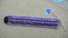
Spirals everywhere! This spiral rope uses purple 4 x 4 cubes, size 6 lavender core beads and size 11 teal accent seed beads to demonstrate the colors in the GMS Fungi Stasin for Candida Albicans, Candida Albicans is a fungal infection caused by yeast.  The colors are so pretty together.

Friday, November 18, 2011

Luxol Fast Blue Bracelet

Luxol Fast Blue is used in the demonstration of pathologic processes in the nervous system. In particular myelin, the white fatty nonliving material, which insulates and protects the nerve fibers are demonstrated. 
This bracelet is a tennis bracelet made Swarovski 4mm crystals  and pladium delica's for the setting with a small Sterling Silver toggle.

Friday, November 4, 2011

Earrings

These are some of my favorite earrings. I especially like the Siam Red dangles with the sterling silver cones. I call them my Congo Red earrings. They have a silver flower between two Swarovski Siam 4mm bicones crystals. I hope you all are planning some fun projects for the weekend. I have several orders to fill.

Saturday, September 3, 2011

Von Kossa Calcium Stain Bracelet

Calcium salts are normally present in bones and teeth. Abnormal deposits may be found in areas of TB infection, lymph nodes, fibroids, etc. This Von Kossa Calcium Stain bracelet depicts the brown to black calcium salts  using iris 3 x 3 mm cubes surrounded on all sides by iris seed beads with their various shades depicting the pink to red cytoplasm and nuceli. In the lab, tissue sections are treated with a silver nitrate solution and silver is deposited replacing the calcium. It is then reduced by a strong light and is visulized as metallic silver. That is why I picked the metallic iris cubes used in this bracelet.  This bracelet lays flat against your wrist making it a good choice for daily wear.

Congo Red Amyloid Bracelet

This stunning tennis bracelet depicts the Congo Red Amyloid Stain. The Congo Red Amyloid Stain demonstrates myeloma-associated or tumor-associated amyloidosis.  Amyloid deposits may become very large and damage surrounding tissue.  This bracelet uses 4mm Siam Swarvoski Crystals surrounded by Palladium Plated Miyuki Delicas in a single row. It looks fabulous with your evening wear or stunning with jeans and a tee-Shirt.

Elastic Stain Necklace

This beautiful necklace depicts the Elastic Stain. The Elastic Stain demonstrates atrophy, arteriosclerotic changes and helps the pathologist to determine whether blood vessels have been invaded by a cancer. Black Iris 4 x 4 cubes are used to depict the black elastic fibers which Verhoeff's working solution stains and iris seed beads depict the pinks and red collagen that is counterstained byVan Gieson's.

Wednesday, August 3, 2011

Bile Stain Bracelet

This is my Bile Stain Bracelet. In it I have made a herringbone pattern on both ends in green 4 x4 cubes with 11* Seed Beads. In the center of the bracelet I put rows of alternating, pink crystals, pink 8* Seed Beads in a ladder stitch,and light brown crystals. I made a seed bead toggle clasp.

The Bile Stain demonstrates bile pigments in tissue. Biliary obstruction may cause excessive amounts of bile pigments in the liver. When red cells ( a component of blood ) degrade it results in a normal production of Bilirubin. In the stain, the tissue colors range from olive green to emerald green depending on the concentration of bile pigment present. Muscle and cell cytoplasm stains yellow and collagen stains pink. So, the colors in my Bile Stain Bracelet demonstrate how bile would show up in the tissue.

Tuesday, July 5, 2011

PAS Stain Bracelet

There are many uses for the PAS stain. One of the most common is the demonstration of fungus as well as the demonstration of mucosubstances secreted by epithelia of various organs, such as the disgestive tract, lungs, and cervix. Glycogen, normally found in many cells, skin, liver, muscle is also demonstrated by the PAS stain.  It is important to show an increase in the amount of glycogen present in certain pathologic conditions.  Certain types of cancer show an increase in glycogen. There are various methods for the PAS. Most show pink to red basement membrance, fungi, glycogen and mucin. A light green or purple (hematoxylin) counterstain can be used.

I had a great time designing this bracelet, since it is one of my favorite stains. I do quite a few of them, mostly looking for fungus. I used the light pink cubes as the base of the bracelet in a ladder stitch, some fuchsia and raspberry seed beads with satin peridot Swarovski crystal in an X shape as the center. I just love the satin peridot. I think it is my new favorite color. I also love the way the design has an openness to it.

Friday, June 17, 2011

Award Winning Designers

Oh my goodness, I really messed up big time. My sister, Linda and her group came in second on the Swarovski Challenge during their cruise not third! I am even more impressed!  Here is the link to the Swarovski website with their winning design pictured. Take a look. It is  truly beautiful. http://www.create-your-style.com/Content.Node/events-and-stores/events/anchors_away_Cruise_2010.en.php#Winning